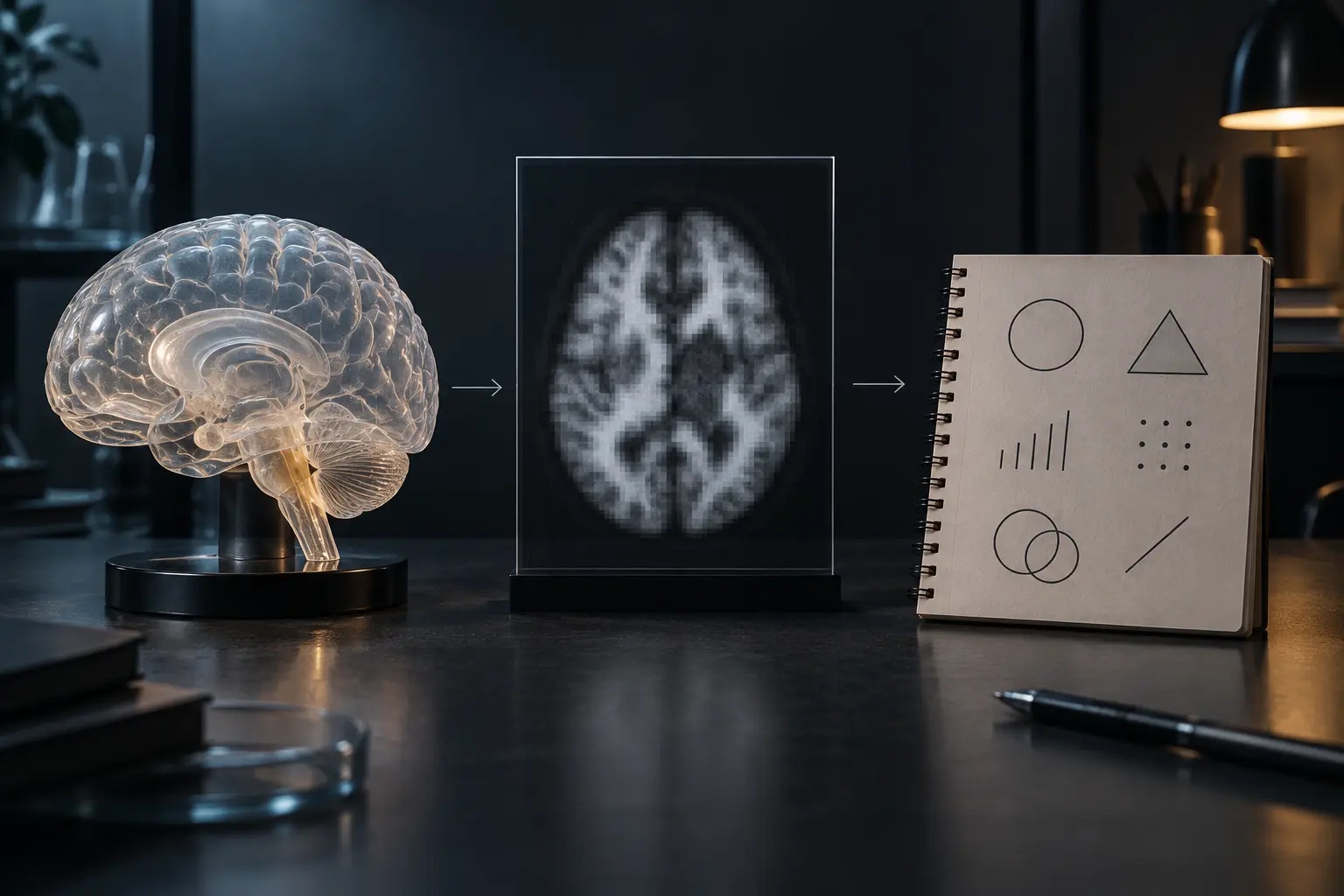
A brain image can be anatomically detailed, statistically sophisticated and genuinely useful without being a photograph of a thought. That distinction matters because the picture arrives at the reader after a chain of measurement, preprocessing, modeling, comparison and visualization. The final map may support a careful claim about a signal under specified conditions. It does not let an observer look directly at fear, belief, intention or consciousness.
The temptation is understandable. A brain-shaped image looks more immediate than a table of coefficients. Bright regions look like locations where a mental state has been found. But visual immediacy is not evidential immediacy. The map is the last interface in a long measurement pipeline.
What functional MRI measures
Structural MRI and functional MRI answer different questions. Structural imaging can reveal anatomy. Common functional MRI methods use the blood-oxygen-level-dependent signal, usually called BOLD. Neural activity changes local energy demand; vascular responses alter blood oxygenation and magnetic properties; the scanner detects signal differences related to that response. The relationship is valuable, but indirect.
The National Institute of Biomedical Imaging and Bioengineering's MRI overview describes functional MRI as a method that maps activity by detecting changes in blood flow. That sentence contains the discipline the popular image often loses: activity is inferred through a hemodynamic proxy. The proxy has timing, spatial and physiological constraints. Movement, breathing, scanner behavior and analysis choices can affect the observed pattern.
Indirect does not mean fake. A thermometer is indirect. So is a blood-pressure cuff. Science is full of instruments that turn an inaccessible process into a measurable signal. The correct response is not dismissal. It is to keep the measurement model attached to the conclusion.
The image is a pipeline, not a window
Raw scanner data do not arrive as the clean activation map seen in an article. Researchers correct for motion, align images, define regions or whole-brain comparisons, smooth or transform data, choose statistical models and set thresholds. Each step can be justified. Each also carries assumptions.
A responsible reading asks which contrast produced the image. “Activity during memory” is too broad. Was the comparison recalling words versus reading them, correct trials versus errors, one group versus another, or a model prediction against baseline? A colored region means little without the task, comparison, sample and analysis.
The same caution applies to absence. A region that does not cross a reporting threshold has not been proven inactive. The study may lack power, the signal may be noisy, the timing may miss the event or the analysis may distribute the effect across a network. A map is a result under a method, not an inventory of everything the brain did.
Thresholds also shape the visual story. A continuous statistical surface is often converted into a simpler image that highlights values above a chosen cutoff. That makes the result legible, but the boundary between colored and uncolored tissue is not necessarily a biological border. Different correction methods, regions of interest and display choices can change which parts command attention. The paper's methods and numerical results carry more information than the thumbnail used in a headline.
Ask whether the researchers specified their analysis before seeing the result, whether they tested the finding on held-out data and whether the effect appeared in an independent sample. Exploration is useful for discovering patterns; confirmation is a different job. A result found and evaluated on the same data can be a good hypothesis while remaining weak evidence of generalization.
The reverse-inference trap
Suppose a region is more active during a task associated with anxiety. It does not follow that activity in that region, observed elsewhere, proves anxiety. Brain regions and networks participate in multiple processes. Moving from task to signal is already an inference; moving backward from signal to one mental state adds another.
This is the same error exposed by the claim-evidence-inference protocol: evidence can be compatible with several explanations. A strong analysis names plausible alternatives and shows what would distinguish them. A weak one treats a familiar label as if it were the only cause.
Researchers sometimes use multivariate patterns or predictive models rather than single-region stories. Those methods can improve classification under tested conditions. They do not remove the need to specify the population, task, scanner, preprocessing and validation boundary.
Decoding is not mind reading
“Brain decoding” can mean that a model predicts which of a limited set of stimuli or tasks best matches a measured pattern. Performance may be impressive. But choosing among categories defined by the experiment is not the same as reading unrestricted thought. The model is trained on examples, a response format and a measurement environment. It succeeds inside that frame.
Generalization is the hard part. Does the model work on new people, a different scanner, ordinary life, a changed task or a person who deliberately uses another strategy? A classifier can exploit a stable correlate without capturing the mechanism researchers care about. A model can be right for the wrong reason, whether the input is text, pixels or brain signals.
Subjective experience adds a further boundary. Correlates of reported experience can constrain theories of consciousness. They do not convert first-person experience into a third-person image. The relation between report, behavior, neural process and experience remains a scientific and philosophical problem—not a gap that color alone closes.
A group result is not automatically an individual diagnosis
Many studies estimate average differences across groups. An average pattern can coexist with substantial overlap among individuals. It may describe a sample while remaining unreliable for classifying one person. Clinical use requires validation for the intended population, setting and decision, not merely a statistically significant research contrast.
Individual interpretation also raises consequences. A finding used to guide surgery, support a diagnosis or make a legal claim needs stronger evidence than a finding used to explore a hypothesis. The cost of error changes the required validation.
Base rates matter too. Even a model with apparently strong sensitivity and specificity may produce many false positives when the target condition is rare. Readers should ask for absolute numbers, the prevalence in the tested population and the decision threshold—not just a single accuracy figure. The same pattern can be useful for group research yet unsuitable for screening an individual.
The brain-image reading card
| Question | What to locate | What it prevents |
|---|---|---|
| What was measured? | Structure, BOLD, blood flow, electrical activity or another signal | Treating every brain image as the same evidence |
| What was compared? | Task, baseline, group, time point and contrast | Reading color without its experimental condition |
| What was inferred? | The exact mental or biological claim | Turning association into direct observation |
| What else fits? | Movement, strategy, arousal, physiology and alternative cognitive processes | Reverse inference and single-story explanation |
| Where was it validated? | New participants, sites, scanners and real-world settings | Confusing in-sample fit with general use |
| What decision follows? | Exploration, prediction, diagnosis or intervention | Using research evidence beyond its tested purpose |
What brain imaging can establish
None of this reduces brain imaging to decorative neuroscience. Imaging can reveal anatomy, map function before surgery, test relationships among tasks and signals, compare models, follow change and identify patterns worth further study. Its power grows when the claim stays close to what the instrument and design can carry.
The skeptical position is not “the scan means nothing.” It is “state exactly what the scan means here.” Good science survives that demand. Certainty theater does not.
Sources and further reading
- NIBIB: Magnetic Resonance Imaging, for the official overview of structural and functional MRI.
- Logothetis et al., “Neurophysiological investigation of the basis of the fMRI signal”, for a foundational investigation of the relationship between neural activity and BOLD response.
- NCBI Bookshelf: Functional Magnetic Resonance Imaging, for clinical and technical background.
END OF TRANSMISSION 044
Keep the question. Test the model.
Choose the narrowest claim the evidence can carry, then leave room for revision.